The most common form of cancer in the United States is skin cancer. The two most common forms of skin cancer are basal cell carcinoma, and squamous cell carcinoma. Melanoma is the third most common skin cancer which is more severe and causes the most deaths.
About 3 million people in the U.S. are diagnosed with nonmelanoma skin cancer each year, and basal cell carcinoma (BCC) accounts for about 80 percent of these cases, according to estimates.
BCC and squamous cell carcinoma ( SCC) are normal and highly treatable, in comparison to melanoma.
We look at the diagnosis and treatment of those carcinomas in this article.
What is carcinoma?

The two most common types of skin cancer include BCC and SCC.
Carcinomas are also classified as skin cancers but are not melanoma. A carcinoma is a cancerous tumor of the epithelial tissue, which is the tissue underneath the skin.
Also present in the digestive tract, blood vessels, and other organs is epithelial tissue which means that carcinomas can affect areas of the body other than the skin.
BCC is more common than squamous cell type, many times. There is also a rare type of skin cancer called Merkel cell carcinoma.
For the vast majority of cases, individuals who obtain a diagnosis of carcinoma are over 50 years of age. Statistics also indicate that in people with white skin 90 percent of carcinomas occur.
Types
Healthcare professionals define the different carcinomas by the type of cell in which they occur.
Basal cell carcinoma
BCC grows in the basal cells, which are round skin cells that lie far below the squamous cells in the skin’s epidermis. They form the epidermis base layer which meets the dermis.
BCC is unlikely to spread, but doctors who believe that this form of carcinoma is present in an individual will still refer them for further examination.
Squamous cell carcinoma
Squamous cells make up much of the skin’s top layer which people call the epidermis. These cells are flat and scale-like.
Doctors suspecting SCC will provide a more urgent referral, since it is more likely to spread than BCC.
But SCC is much rarer than BCC. It has less than 20 percent responsibility for nonmelanoma skin cancers.
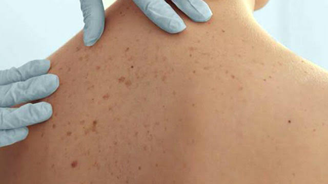
Pictures


Causes
The primary cause of carcinoma and other skin cancers is exposure to ultraviolet ( UV ) radiation from sunlight.
Some people are more sensitive to UV light than others, and are more vulnerable to the cancer development effects of sunlight. For example , increased UV exposure from tanning beds and UV drying lamps in nail salons can add to a person’s risk as well.
UV radiation in skin cells can cause damage to the DNA, resulting in mutations during cell division, and likely leading to skin cancer.
Risk factors
Factors and features that increase the risk of carcinoma include a personal history of skin cancer and radiation treatment for any form of cancer, especially in childhood. A family cancer history can also contribute.
Further risk factors include:
- having numerous, irregular, or large moles or freckles
- a tendency to burn before getting a suntan
- having fair skin, blue or green eyes, or blond, red, or light brown hair
- autoimmune diseases, such as systemic lupus erythematosus (lupus)
- inherited conditions, such as xeroderma pigmentosum and nevoid basal cell carcinoma syndrome, also known as Gorlin syndrome
- a weakened immune system, possibly due to HIV, receiving an organ transplant, or taking immunosuppressant drugs
- taking medicines that make the skin photosensitive, such as vandetanib (Caprelsa), vemurafenib (Zelboraf), and voriconazole (Vfend)
- human papillomavirus (HPV) infection, particularly in people with a weakened immune system
Actinic keratosis, which consists of rough, raised growths causing precancerous changes in skin cells, is an SCC-specific risk factor. The most common form of precancerous skin lesion is those growths.
That condition may develop into skin cancer without treatment.
While UV radiation is the leading risk factor for SCC, the following skin damage may also increase the risk of carcinoma of this type:
- burns to the skin
- chemical damage
- exposure to X-ray radiation
BCC may also occur in childhood after exposure to X-ray radiation, though this is a much less common cause of carcinoma than exposure to UV.
Symptoms
Both BCC and SCC are skin tumors, and have several characteristics in common. Nevertheless, the presence of these skin lesions can differ.
Some carcinomas retain a flat surface and may look like healthy skin as a result. Anyone with any unexpected lesions will contact and track a healthcare provider for a checkup.
Apart from its appearance, a lump or lesion in its early stages sometimes causes no obvious symptoms. It may not be visible as a result until it is fairly big where it may itch, bleed, or cause pain.
Basal cell carcinoma
Usually BCC is presented as a shiny papule, a small red or pink lump which grows slowly.
After some months or years, a shiny, pearly, or waxy-looking border can develop.
A raised edge often rings a central ulcer, and it can become apparent to abnormal-looking blood vessels. These may emerge as blue, brown , or black areas. Instead, they may be pink growths or patches of pale or yellow resembling scars.
Because of this wide range of appearances it is important to receive an accurate diagnosis from a doctor.
BCC can look scaly, sometimes causing frequent crusting or bleeding. This that look like a healing scab as it crusts over but sores may still appear. Individuals with BCC also seek medical help when they come across a sore that is not healing.
Squamous cell carcinoma
Usually SCC is viewed as persistent, thick, rough, scaly patches or as a firm pink lump with a flat, scaly, and crusty surface.
Such lesions can bleed when bumped, scratched, or scraped by a human. Although they often resemble warts, with a crusted surface or raised edge, they can also appear as open sores.
It is important to obtain a health care professional’s advice about the creation of any new growths or improvements in pre-existing skin growths or sores.
Diagnosis

A doctor may do a physical exam to diagnose some type of skin cancer. The skin lesion will be examined and its size, shape , texture and other physical attributes recorded.
You can also take a professional analysis photo of the lesion, or record its current size and appearance for potential comparisons. The doctor will often check up on additional skin symptoms for the rest of the body.
They will also take a medical history that focuses on the lesion and any associated conditions, like sunburn.
Due to their spreading propensity, a doctor must immediately refer suspected SCC cases for specialist examination and treatment. Suspected BCC tumors should not be referred as urgently because they are less likely to spread.
If they believe a lesion may be cancerous, then the doctor is likely to perform a biopsy as well. There are four different forms of skin biopsy all involving the removal of skin tissue for laboratory assessment.
The different types are:
- Shave biopsy: Using a sharp surgical blade, the doctor shaves the top layers of skin cells, usually as far as the dermis but sometimes deeper. This type of biopsy often results in bleeding, but it is possible to stop this by cauterizing the wound.
- Punch biopsy: The doctor uses a sharp, hollow surgical tool that resembles a tiny cookie cutter to remove a circle of skin from below the dermis. A person may need a single stitch to close the resulting wound.
- Incisional biopsy: The doctor removes part of the growth with a scalpel, cutting away a full-thickness wedge or slice of skin. This type of biopsy often needs more than one stitch afterward.
- Excisional biopsy: The doctor removes the whole growth and some surrounding tissue with a scalpel. The resulting wound usually requires stitches.
Once the tissue sample is taken, the doctor will send it under a microscope to a pathology laboratory for analysis. The pathology department must test the cells to detect cancerous traits. If cancer is present, they will determine its type.
For people with BCC, further inquiries are generally not required as this rarely spreads. People with SCC can need to undergo cancer tests in other tissues, however.
Many assessments typically include imagery, and may include:
Staging
When a doctor detects skin cancer, then they may label it as a stage. To do so, they must determine its size and depth and to what degree it has spread to local and remote locations in the body, such as nearby lymph nodes or other organs.
The doctor can also take tissue from lymph nodes close to the carcinoma site to help them stage cancer. A fine needle biopsy is also used by them for laboratory analysis.
Staging may not be performed until after a skin tumor has been surgically removed. The stages range from 0 to 4, with 0 representing in situ carcinoma which only affects the skin’s top layer.
Step 4 carcinoma refers to a carcinoma that has spread across the body to other parts. The stages between describe lesion size, tissue depth, and any nearby invasion.
Treatment

The treatment strategies for both forms of carcinoma are similar, but the medical team puts more focus on monitoring people with SCC for signs of metastasis.
The particular treatment or treatments that the doctor recommends will depend on the size, form, stage, and location of the carcinoma. Additional factors such as potential side effects and individual preference, will also be taken into account by the doctor.
Nonetheless, diagnosis is likely to require a team of health care practitioners including dermatologists and experts in surgical , medical, and radiological cancer.
Possible care choices can include:
Curettage and electrodesiccation: This is a common treatment for a minor lesion removal operation. A small, sharp, spoon- or ring-shaped tool called a curette is used by the doctor to scrape the carcinoma away before burning the site with an electric needle.
A complete removal of the cancer cells may require more than one round of curettage and desiccation.
Surgical excision: The lesion is removed by a surgeon, often in a process called Mohs surgery, which works best with larger lesions. During this procedure, after removing each layer the surgeon tests for the presence of cancer cells.
Mohs surgery is particularly useful in cases that involve removal of as little skin as possible, such as on near-eye lesions. It is also used by doctors on lesions with a high risk of recurrence.
Cryosurgery: This procedure, which involves applying liquid nitrogen to freeze and kill cancer cells, may be used by doctors for small tumours. Then the lesion blisters over and falls off within weeks of treatment.
Topical chemotherapy: The doctor may apply chemicals or medicines directly to the skin which destroy cancer cells.
The option for chemotherapy is 5-fluorouracil which includes Carac, Efudex, Fluoroplex and other drugs. For several weeks a doctor can apply this cancer-killing drug to the skin once or twice a day.
Since this local treatment does not reach other systems in the body, it does not cause side effects for other types of cancer which often occur with chemotherapy.
Nonchemotherapeutic treatment options include the imiquimod cream, sold under the Aldara and Zyclara labels. For small BCCs, this cream is adequate and it works by stimulating the body to produce interferon, which allows the immune system to fight the tumour.
Also, a doctor may directly inject interferon into the lesion.
Radiation therapy: The treatment team is treating lesions with a concentrated radiation that are large or difficult to remove.
Photodynamic Therapy (PDT): often, physicians will use this two-step procedure to treat BCC. They will add a light-sensitive cream to the infected skin region, and then expose it to a powerful source of light. The light has the same wavelength of blue light, which results in the death of carcinoma cells.
Given that the skin remains sensitive to light for the next 48 hours, during this time people should avoid UV light to minimize the risk of severe sunburn.
Laser carcinoma therapy: It includes the use of different forms of laser to kill cancer cells. Many lasers vaporize, or ablate, the top layer of the skin, removing any lesions present in it.
Some lasers are non-ablative and penetrate the skin without the top layer being covered. Their success in treating low, superficial BCCs is some evidence.
BCC laser therapy has not yet been approved by the US Food and Drug Administration ( FDA). However, if other therapies have not been effective, doctors can often use this as a secondary therapy.
Prevention
There is actually no systematic carcinoma screening programme. Instead, people should check for irregular lesions themselves, or ask a doctor for a physical test.
UV light is the primary risk factor for all forms of carcinoma. The best strategy for prevention is to adopt sensible sun exposure practices, and avoid tanning beds.
Minimizing sun exposure: People can reduce their risk of sunburn, skin damage and all types of skin cancer, including carcinoma, by reducing their exposure to UV light.
Although some exposure to the sun is required to maintain adequate levels of vitamin D, which is essential to maintaining skin health, sunburn increases the risk of carcinoma.
People can limit sun exposure by finding shade, usually by 10 a.m., when the sun is at its peak. And, at 4 p.m.
Clothing: Skin-protecting clothes from the sun involve hats with a large brim or crown, sleeve tops and sunglasses.
Clothes in sun-protective fabrics would have UV 400 or UV protection factor (UPF) labels on them. Choose tight weave over loose-weave fabrics for better protection.
When buying sunglasses, check the labels for a 100 percent mention of UVA and UVB radiation protection.
Approved wide-spectrum sunscreens: Pick an effective sunscreen and apply it liberally and consistently to the skin to minimize UV light exposure.
Check the label to ensure the sunscreen protects against UVA radiation and UVB radiation.
Because some sunscreens are ineffective and contain suspected cancer-causing substances, check consumer reports to make sure a particular sunscreen brand is safe and effective prior to use.
Using a sunscreen with a minimum sun protection factor ( SPF) of 30 and reapply it every 2 hours on all exposed skin. Increasing the application after intense sweating or swimming to once an hour. There are also lotions which are waterproof.
Children and youngsters are especially vulnerable to sun exposure. People should also be aware that the levels of UV light are more dangerous at higher altitudes, in places closer to the equator, and at sunny locations throughout the year.
The U.S. Task Force on Preventive Services recommends that infants, teenagers and young adults between the ages of 10 and 24 with fair skin will limit their exposure to UV radiation.
Avoid tanning beds: tanning beds, tanning parlors and sunlamps raise the risk of carcinoma significantly.
Artificial tanning, since it exposes the body to a concentrated source of UV rays, is more harmful than normal sunbathing. Avoid using nail lamps when receiving manicure or pedicure as this may also increase skin cancer risk.
This advice is especially important for people undergoing routine nail therapy.
Self-examination

The fundamental concept of carcinoma screening and other types of skin cancer is to look for unresolved skin changes.
To be successful the skin should be self-examined:
- paying particular attention to areas of skin that get lots of sun exposure
- asking a partner or family member to check difficult-to-see areas and using full-length and hand mirrors
- knowing your skin and learning how moles and marks usually look to recognize any changes
- taking photos, which can help track changes
- checking for changes in size, shape, color, or texture
- performing self-examination in good lighting
- seeking medical attention for any sores that do not heal
- working across the body systematically from head to toe to examine all areas
- checking all areas of the body, including the more intimate ones
- keeping a note of any observations and recording the dates of self-examinations
Those interventions can help individuals detect carcinomas early on and treat them before spreading.
Outlook
Treatment is likely to be more successful in situations where an patient early on detects skin changes and receives prompt medical attention.
Prompt treatment significantly increases the odds of survival in cases where cancer is responsible for changes in the skin and decreases the risk of severe tissue damage and disfigurement.
BCC has an excellent survival rate, since it spreads very rarely beyond the original site. Often, doctors may treat it in-office.
In its early stages, SCC is treatable, and most therapies are more than 90 percent successful. Mohs surgery is the most effective option for 97 per cent of people who receive this treatment to resolve SCC.
However, if SCC extends beyond the original site and enters other body systems, the survival rate would decrease to around 30 percent.
Effective recognition is important for a person’s outlook to be enhanced.
Question:
How do I tell the difference between melanoma and carcinoma?
Answer:
Melanoma commonly occurs in the skin that is regularly exposed to sunlight, like that on the face , neck, hands , and arms.
This is also noticeable, as it makes a skin area look darker than the rest. Melanoma has this effect as it starts in cells called melanocytes which are in the lowest epidermis layer and play a role in the color of the skin.
Nonmelanoma, or BCC and SCC, are respectively in the mid and upper layers of the epidermis. Possible signs and symptoms of these carcinomas include irregular changes in skin appearance, such as ruggedness, redness, scaliness, or thin, high, smooth or red areas.
If you notice anything unusual on your skin, please visit your dermatologist for a physical examination.


