MRI and other advances in medical imaging have increasingly enabled arthritis to be detected in its early stages and its severity assessed.
MRI scans are a powerful tool for arthritis diagnosis, because they are more reliable than traditional X-rays and ultrasound imaging.
MRI is more costly than some other imaging tests, so some hospitals may use it only in clinical trials or for specific conditions, such as spondyloarthritis, to determine.
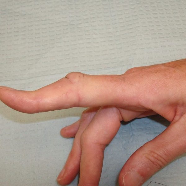
Arthritis is a disease in the joints that causes pain and inflammation. Early detection of arthritis will help people get access to effective treatment faster and delay disease progression.
Read more about what looks like arthritis on an MRI scan, and what to expect during the operation, in this article.
What does it look like?
Arthritis affects mostly the joints and surrounding tissues. Some harm will be noticeable on an MRI scan at these areas.
For the following symptoms of arthritis, a radiologist or other form of doctor should look:
- damage to the cartilage
- bone spurs (osteophytes)
- tears in the ligaments
- an increase in bone density
- synovitis, or inflammation of the synovial membrane in joints
- bone marrow edema, which is a build-up of fluid in the bone marrow
- joint effusion, commonly called ‘water on the knee’
A CT scan may be suitable for bone density assessment. A doctor may use a DEXA scan or a normal X-ray, too. .
Although bone spurs appear on an MRI scan, X-ray images are the best way to detect them. MRI’s are at looking at bone than soft tissue.
MRI scans are highly sensitive compared to other medical imaging techniques and provide detailed pictures. They may represent both the bone and surrounding tissues when looking at a joint.
MRI may suggest that bone loss has occurred, which can occur later on in the disease. It can also tell when bits of tiny bones have broken off.
MRI can also provide detailed pictures of the surrounding tissue, which allows doctors to spot inflammation in soft tissues much faster than when using an X-ray.
MRI can identify bone erosions and assess how much arthritis wears away the cartilage lining the joint before an X-ray shows this damage.
Two of the most common arthritis types a doctor may seek using MRI are:
- Osteoarthritis: A condition in which the tissue in the joints that supports the bones breaks down. This usually occurs with age or from a previous joint injury, causing the joints to rub against each other.
- Rheumatoid arthritis: an inflammatory disease that causes synovial tissue inflammation lining the joints. Autoimmune diseases arise when the immune system attacks its own tissues and parts of the body in the wrong way.
Osteoarthritis vs. rheumatoid arthritis
Other symptoms of osteoarthritis can be clearly identified by MRI, including whether cartilage is wearing away.
MRI may also detect symptoms of rheumatoid arthritis, but a doctor will also use a number of other tests, for example blood tests.
Using MRI, doctors can make a distinction between soft tissues and fluids. This means they can identify signs of rheumatoid arthritis, such as inflammation and the synovial membrane disease.
The synovial membrane protects and guards joints. For people with rheumatoid arthritis it may be swollen (synovial thickening).
What to expect
Having an MRI scan is a very safe procedure in most people, with few side effects.
People with an MRI scan typically do not need to make any special preparations, although they may need to change into a gown in the hospital.
Because MRI machines use powerful magnets, removing any metal objects, such as watches or jewelry, is necessary prior to scanning.
The MRI scanner is a large machine, with a donut shape. The person who has the scan must lie down in the middle of the scanner on a bed. Depending on what part of the body needs scanning the exact position will vary.
Before starting the scan, a radiologist or technician can explain the procedure and any information regarding safety. The scan time can be between 45—60 minutes per part of the body everywhere.
It is important to stay as quiet as possible during the scan. While it is working, the scanner will make loud noises which could make some people feel uncomfortable or anxious. Technicians could give earplugs to help block that out, or a person may listen to music.
Inside the scanner, there is a two-way intercom which a person can use if they have any questions or problems with their procedure. An MRI scan can always be stopped at any given point. This can be done inside the scanner via an alarm button or by using the intercom.
People with claustrophobia or who consider the experience very uncomfortable may have the option to take a sedative or use an open-MRI machine during the testing.
Most people, like the ones with implanted pacemakers, can not have an MRI scan.
Other diagnostic tests
During a diagnosis a doctor will always determine a person’s physical symptoms and medical history. They can record temperature, heart rate, and/or gland swelling.
In some cases, it will take medical imaging to assess the damage or confirm the diagnosis. Although MRI is one alternative, X-rays will reveal bone spurs and whether the joints have become too close to each other.
Another possible test would be a CT scan. These aid in the detection of bone lesions.
Occasionally, a doctor may use an ultrasonic scan to detect synovial membrane or tendon problems.
When to see a doctor
Lesser aches or pains are common around the joints. Over age or after a vigorous exercise this becomes more normal.
But if joint pain lasts for several days, comes over swelling, or makes daily activities difficult to do, it’s best to see a doctor.
Summary
MRI scans are of great help in the procedure of arthritis. They can provide detailed images of the tissues around and in the joint.
These videos are helping physicians make a diagnosis and determine the seriousness of the disease.
For arthritis, having an MRI scan is generally a safe treatment. Physicians may also recommend other examinations, such as X-rays and ultrasounds, in some cases.